How to Tell Acne Marks Apart from Pigmentation and Dark Spots
Red acne marks and brown acne marks, melasma, freckles and dark spots — learn how they form differently by skin layer, how to tell a colored mark from a pitted scar, how lasers work by target, and what aftercare actually matters.
Written by ATTITUDE Clinic Editorial Team
Some people find that after acne settles, the spot stays pink. Others are left with a brown mark. Others notice a slight dip when they touch it. They all get called "acne marks," but what happened inside the skin is not the same. So are these marks the same problem as the dark spots people usually talk about? What separates a red mark from a brown one, how long is it reasonable to wait, and in what order should you approach it? Let's set out the criteria one by one.
The short answer first
The color of an acne mark comes from one of two directions. A pink or red mark comes from blood vessel changes left behind where inflammation has passed, while a brown mark is the result of increased melanin pigment in the same spot. If the skin surface is genuinely indented, it is not color but a scar with damaged dermal structure, so the approach differs from treatment aimed only at pigment. Melasma, freckles and dark spots also start from melanin, but which layer they settled in and why differs from case to case, so using the same device at the same intensity is difficult. That is why the order is always the same. Before erasing color, confirm which layer the remaining mark belongs to. Even for the same mark, the response splits depending on how strong the inflammation was, how much UV exposure there was, and the skin tone and barrier condition.
Why do red marks and brown marks split apart?
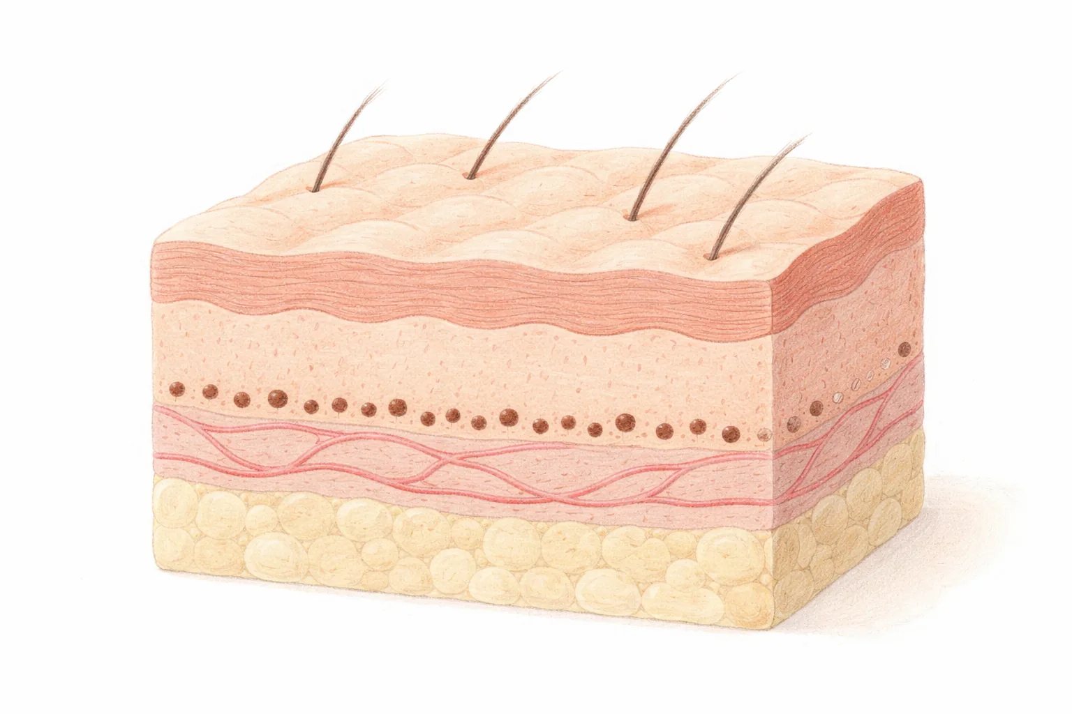
Skin color is determined by the amount of melanin, and melanin is produced by melanocytes scattered in the basal layer, the deepest layer of the epidermis (MSD Manuals, overview of skin pigmentation). Hyperpigmentation can appear from a range of causes, including inflammatory conditions, the inflammatory state of healed skin, and sun exposure. Brown marks after acne sit on this same continuum. Melanin is produced in greater amounts to protect the skin when it is stimulated by UV, heat, inflammation or hormonal change, and if the inflammation was strong and long-lasting, or acne repeated in the same spot, that much more accumulates (Melasma, freckles, dark spots, sun spots — are they all the same?).
Red marks come from the blood vessel side, not the pigment side. While inflammation is settling, dilated superficial vessels overlap with the recovery process, which is why the area looks pink. That is why lasers aimed at pigment or whitening ingredients alone do not respond well to redness, and the approach itself has to be set up differently. It is also not uncommon for redness and brown color to overlap in the same area. Because color looks different depending on lighting and skin tone, it is hard to divide causes with one photo or one color.
In the end, what determines the depth and duration of a brown mark is the intensity and repetition of inflammation, and the UV exposure that follows. When a red mark fades depends on how often new inflammation rises in that spot. If irritation continues, new marks appear faster than old ones fade.
How is a mark that keeps its color different from a pitted scar?
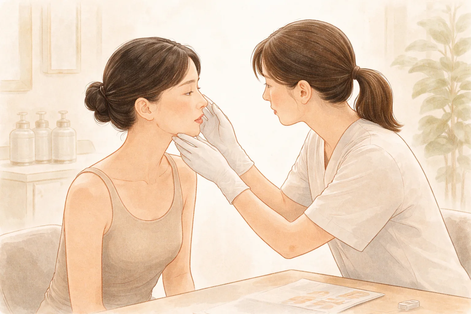
A mark is a state where only color remains without much change in surface height. A pitted scar, by contrast, is a state where dermal collagen has decreased and the surface is genuinely indented, and the contour remains even after the color fades. This is why the cause differs depending on whether it is the color of an acne mark or a pitted scar, and the approach differs too (Does frequent laser treatment really thin the skin?). In consultation, we shine light across the area to look at surface height and shadow together.
How lasers handle these two also differs. For pigment, the approach is to have melanin selectively absorb light of a specific wavelength and break it into fine particles, while for scars and pores the approach is to deliver very fine column-shaped micro-stimulation into the skin to induce regeneration. The latter delivers energy to only some of the openings like a sieve and leaves the rest intact, so the undamaged surrounding cells are helped to build new collagen (Dark spots, redness, scars — can lasers do it all?).
Scars split further depending on shape. Ice-pick or boxcar scars that are deeply indented are hard to fully improve with fractional laser alone, so filler may be used to lift the base or subcision (a procedure that releases the tethering under the scar) may be combined. Raised hypertrophic scars may be better suited to steroid injections or pressure therapy than laser. Even scars with the same name need their shape confirmed first.
Can acne marks be called dark spots too?
Melasma, freckles and dark spots all start from melanocytes, the cells that make pigment. What differs is where, why, and how deeply they settled. Dark spots are pigment patches caused by cumulative UV exposure and aging, with relatively clear borders and often concentrated in the epidermal layer. Freckles have a strong genetic predisposition and repeat a pattern of darkening with UV exposure and fading when UV is blocked. Melasma involves hormones, UV and heat stimulation acting together, leaving pigment cells overactive with indistinct borders that spread widely, and it is divided into epidermal, dermal and mixed types. Sun spots are not accumulated pigment but a benign lesion in which epidermal cells thicken, so the principle is different from the start (Melasma, freckles, dark spots, sun spots — are they all the same?).
By this standard, a brown mark after acne is one branch of pigmentation, but its trigger differs from dark spots. The difference is that the trigger was inflammation rather than cumulative UV exposure, and that the distribution and depth of the spot follow the area where acne occurred. So lumping them under the single word "dark spots" and approaching them with the same settings tends to produce mismatched results. American Academy of Dermatology materials also advise that treating pigment marks begins with understanding why that color appeared (AAD, Age spots and dark marks).
One point to keep in mind. A mole that is new or growing, or one whose shape or color changes or that itches or bleeds, should be checked by a doctor for skin cancer before any cosmetic procedure. Some skin cancers can look like an ordinary brown spot.
What does a laser do for each cause of a mark?
A laser uses heat, but it is not a device that burns the whole skin. Wavelength and intensity are adjusted so that energy is absorbed only by a specific target such as pigment, blood vessels or water, and this is called selective photothermolysis — the principle of concentrating energy on the target while touching the surrounding normal skin less (Does frequent laser treatment really thin the skin?). Because the target that responds differs by wavelength, a laser aimed at pigment and a laser aimed at red blood vessels use different devices.
A picosecond laser has a pulse duration as short as one trillionth of a second, so heat spreads to the surroundings relatively less, and it breaks pigment particles into fine pieces for the body to clear gradually. However, it is hard to expect improvement of vascular concerns with a picosecond laser, and red marks are approached with the vascular laser family, which uses wavelengths absorbed by oxygen-carrying hemoglobin (Dark spots, redness, scars — can lasers do it all?).
Laser toning and dark spot lasers also have different purposes. A dark spot laser concentrates on individual patches and approaches with a short wavelength such as 532nm pico, while toning repeats low-energy 1064nm over widely spread pigment to even out skin tone. If strong energy is used all at once on widely spread pigment like melasma, the pigment-producing cells can be stimulated and actually darken, causing rebound pigmentation, so the basic approach is to lower the intensity and repeat (Melasma, freckles, dark spots, sun spots — are they all the same?). Sometimes the two methods are combined on the same day, and sometimes only one is focused on depending on the concern.
Table 1 What does a laser do for each cause of a mark?
Category
Layer or cause
Usual approach
What to expect and check
Red mark (post-inflammatory erythema)
Superficial blood vessel changes
Vascular laser family (Vbeam)
Redness and swelling for a few days, bruising takes 1–2 weeks to fade
Brown mark (post-inflammatory hyperpigmentation)
Increased melanin
Pigment laser (Picoplus) and UV protection
Low-power repetition is the default, can darken again with UV
Pitted scar
Loss of dermal collagen
Fractional laser, filler or subcision combined if needed
4–6 sessions is a common example, 4–8 week intervals
Dark spots and freckles
Melanin in the epidermal basal layer
Pico spot (532nm pico)
1–3 sessions is a common example, 4–8 week intervals
Melasma
Mixed epidermal to dermal
Low-energy repeated pico toning (1064nm) plus UV blocker
8–12 or more sessions is a common example, at least 1 week apart
Swipe sideways to see the whole table.
In short even on the same face, redness comes from blood vessels, brown color from melanin, and indented spots from skin structure, so the approach splits. Before asking "which laser is good," confirming "which side my mark came from" is the right order. If several causes overlap, dividing priorities rather than doing everything at once reduces the burden on the skin.
If this is your situation
If a wedding or shoot date is already set
Recovery time stretches from a few days to 1–2 weeks depending on the type and intensity of the procedure. Counting backward from your schedule, the type, intensity and start timing can be adjusted, so it helps to tell us the date first. There is also the option of matching your schedule with low-power maintenance rather than rushing a high-power procedure earlier.
If you want steady management
Pigment is not something you erase once and finish. Without UV protection to back it up, it rises again in the same spot. Dark spots are treated at 4–8 week intervals, and melasma toning at least 1 week apart, checking the response as you go. Setting what is needed and in what order together can keep you from needlessly increasing the number of sessions.
If your skin is sensitive or reddens easily
When the barrier is weak, the same settings can be a burden. If cosmetics sting or redness lingers, it is better to mention this before the procedure so the intensity can be set to suit your skin. Repeating before recovery is complete can weaken the skin barrier, leaving it sensitive or red for longer (Does frequent laser treatment really thin the skin?). Adding exfoliation and whitening ingredients all at once in the same period also increases irritation.
If you are a short-stay international visitor
Your length of stay and your outdoor plans after returning home need to be looked at together. High-power procedures with long downtime are too much for a short schedule, while low-power toning or maintenance-purpose procedures are relatively light. Whether you can keep up UV protection after the procedure also belongs in the plan.
Please tell us before your consultation — your recent UV exposure and outdoor schedule, medications you take or apply, exfoliating or whitening ingredients you are using, any experience of pigment darkening after a previous procedure, pregnancy or breastfeeding status, and fixed dates such as a wedding or a shoot. This information is used to set the settings and the order.
How ATTITUDE Clinic proceeds
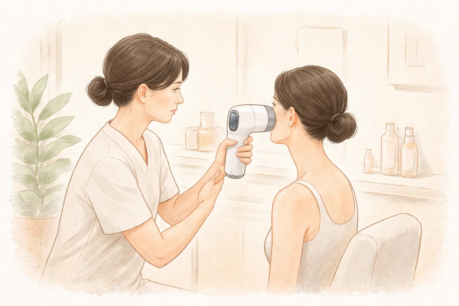
AI skin analysis record — We record and organize current state such as pigment, redness, skin texture and pores to support the consultation. The analysis tool records and organizes information and does not make a medical diagnosis.
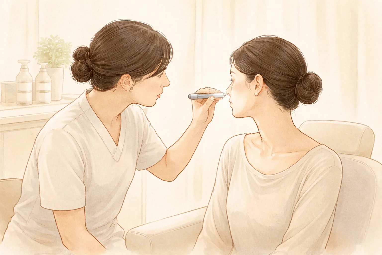
Doctor consultation and type assessment — Adding your UV exposure, lifestyle habits and concerns to the record, the doctor examines your skin directly and judges whether redness, pigment or contour is the main issue. What is needed, what is not, and the order are also decided here.
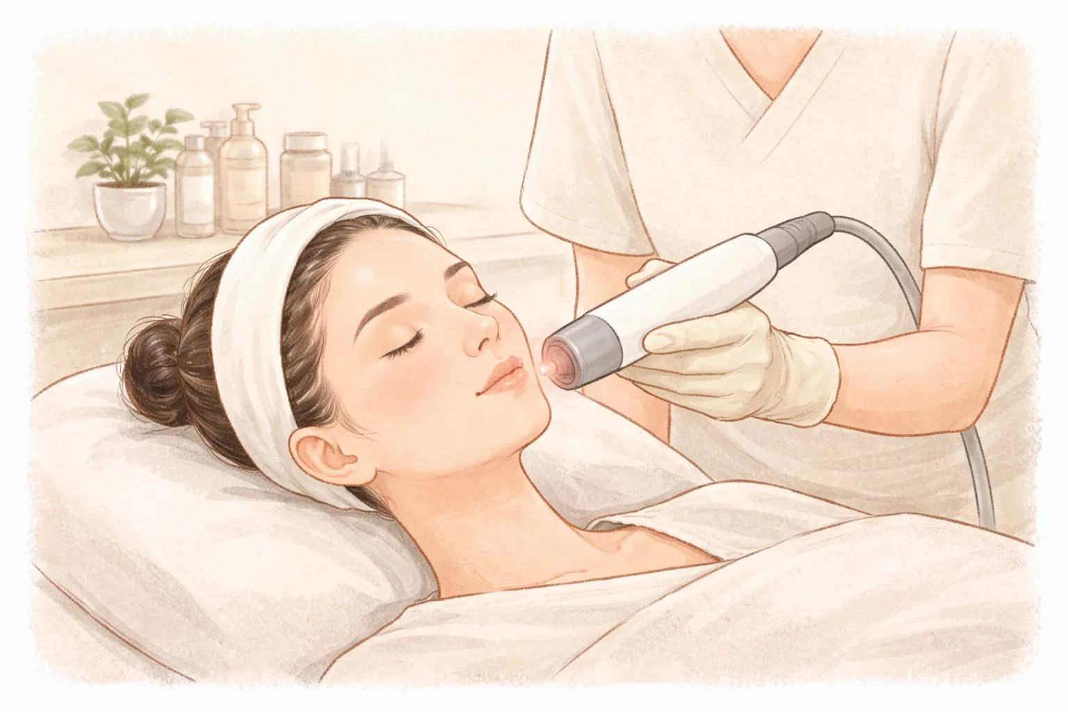
Procedure performed by the doctor — Among the devices we hold, including Picoplus, Vbeam and erbium YAG laser, the device that matches the cause is chosen and the doctor adjusts intensity and combination.
Recovery and follow-up check — Watching your response after the procedure, we adjust the interval and intensity of the next session. We base decisions on actual skin response rather than filling a predetermined number of sessions.
Aftercare and side effects
Calming — Right after the procedure, skin temperature is elevated. Avoid saunas, jjimjilbangs and intense exercise for 1–2 weeks, and wash lightly with lukewarm water without rubbing.
Moisturizing — With the barrier temporarily weakened, keep up moisturizing. Pause exfoliation and scrubs for the period you are advised.
UV protection — Apply SPF 50+ sunscreen for at least 2 weeks and reapply every 2 hours outdoors. If protection is neglected during this period, pigment can settle in the newly forming skin and undo the effect of the procedure (Dark spots, redness, scars — can lasers do it all?).
Follow-up check — Do not pick off scabs; leave them until they fall off on their own. If redness or stinging lasts more than 3 days or suddenly worsens, do not endure it — get it checked.
Common reactions are redness or swelling at the treated area, scabbing, or temporary darkening of color. Vbeam can cause bruising, and redness and swelling last a few days while bruising takes 1–2 weeks to fade. Small scabs after a dark spot laser usually fall off in 7–10 days. Rarely, hypopigmentation (lightening of skin color), blisters or scars can occur, and pigment can rise again over time. These changes are explained by the doctor before the procedure along with the plan.
If redness or heat lasts longer than expected or pain worsens, it is better to come in and get it checked rather than waiting for your next appointment.
Frequently asked questions
Do acne marks count as dark spots?
A brown mark with remaining pigment is, broadly speaking, one branch of pigmentation. However, the trigger was inflammation rather than cumulative UV exposure, and the spot follows the area where acne occurred, so the trigger differs from what people usually call dark spots. That is why, rather than approaching it with the same settings as dark spots, when and after what kind of inflammation it appeared should be looked at together. Red marks are a different branch entirely, caused by blood vessels.
How does the management of red marks and brown marks differ?
For red marks, the approach centers on targeting blood vessels, and lasers aimed at pigment are unlikely to produce a response. Brown marks are handled on the melanin side, so the default is repeating at low power or watching the course along with UV protection. If the two overlap in one area, the target is divided per area and an order is set, and vascular and pigment procedures may be combined on the same day. Which is the main issue is judged by examination.
Can I tell them apart at home just by color?
By color and location alone, it is difficult. The same mark looks different under different lighting and on different skin tones, and it is not uncommon for redness and brown color to overlap or for contour changes to be present as well. Taking photos at intervals from the same spot, same lighting and same distance helps with comparing changes. Dividing the causes requires an examination.
How many sessions does mark treatment take?
Looking at common examples, dark spots are 1–3 sessions at 4–8 week intervals, melasma toning is 8–12 or more sessions at least 1 week apart, and acne scars treated with fractional laser are 4–6 sessions at 4–8 week intervals (Dark spots, redness, scars — can lasers do it all?). These are only common examples and vary with pigment depth, skin condition and degree of UV exposure. Rather than fixing the number of sessions in advance, adjusting interval and intensity while watching the response is safer.
How should I choose a clinic for mark treatment?
There are a few things to check rather than a device list. Does the clinic first distinguish whether your mark is pigment, vascular or contour before the procedure, does it explain by dividing the target per area, and does it cover pain, recovery time and aftercare precautions during the consultation. It is also good to check whether you can get things checked if discomfort arises after the procedure. Even with the same device, how the settings and order are set divides the results.
At a glance
Table 2 At a glance
Item
Key point
Red mark
Blood vessel changes left after inflammation, approach targets blood vessels
Brown mark
Increased melanin after inflammation, UV protection and low-power repetition are the default
Pitted scar
Loss of dermal collagen, approach differs from pigment treatment and may be combined depending on shape
Dark spots and melasma
Same melanin family but different layer and cause, so settings split
Order of confirmation
Judge which layer is involved before color, AI skin analysis record then doctor examination
After the procedure
Do not pick scabs, SPF 50+ UV protection for at least 2 weeks
This article offers general health information. It is not a substitute for diagnosis or treatment based on your own condition, so please talk through your situation with a doctor.