Pico Laser vs. Melasma and Sun Spots: How Doctors Tell Them Apart
Melasma, sun spots, freckles and seborrheic keratoses all look like brown marks, but the layer the pigment sits in and the cause differ. Here is how doctors tell them apart, why the pico laser approach changes, and what to expect after treatment.
Written by ATTITUDE Clinic Editorial Team
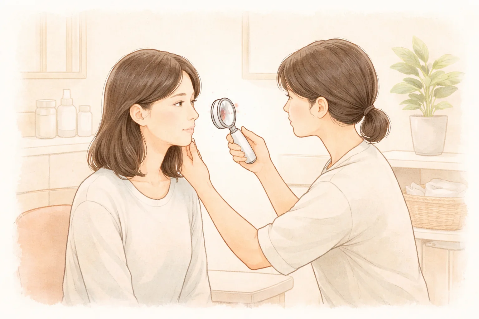
In the clinic, patients often say they can't tell whether the brown patch spreading across their cheek is melasma or sun spots, or whether the raised spot at their temple is a seborrheic keratosis. Different names mean different mechanisms, different depths of pigment, and different treatment directions. Let's walk through how to sort out brown spots and what separates the results.
The bottom line first
Melasma, freckles and sun spots all start from melanocytes — the cells that produce melanin and distribute it to surrounding cells — while seborrheic keratosis is not accumulated pigment but a benign thickening of the outer skin cells, so the principle is different from the start (ATTITUDE Clinic column).
So seborrheic keratosis is usually treated by smoothing away the thickened area with an erbium:YAG laser, whereas melasma involves overactive pigment cells, and using strong energy in one go can actually darken the color. In the end, the criteria for sorting brown spots are not the name but how deep the pigment sits and what triggered it.
Why do they all start from one melanin but have different names?
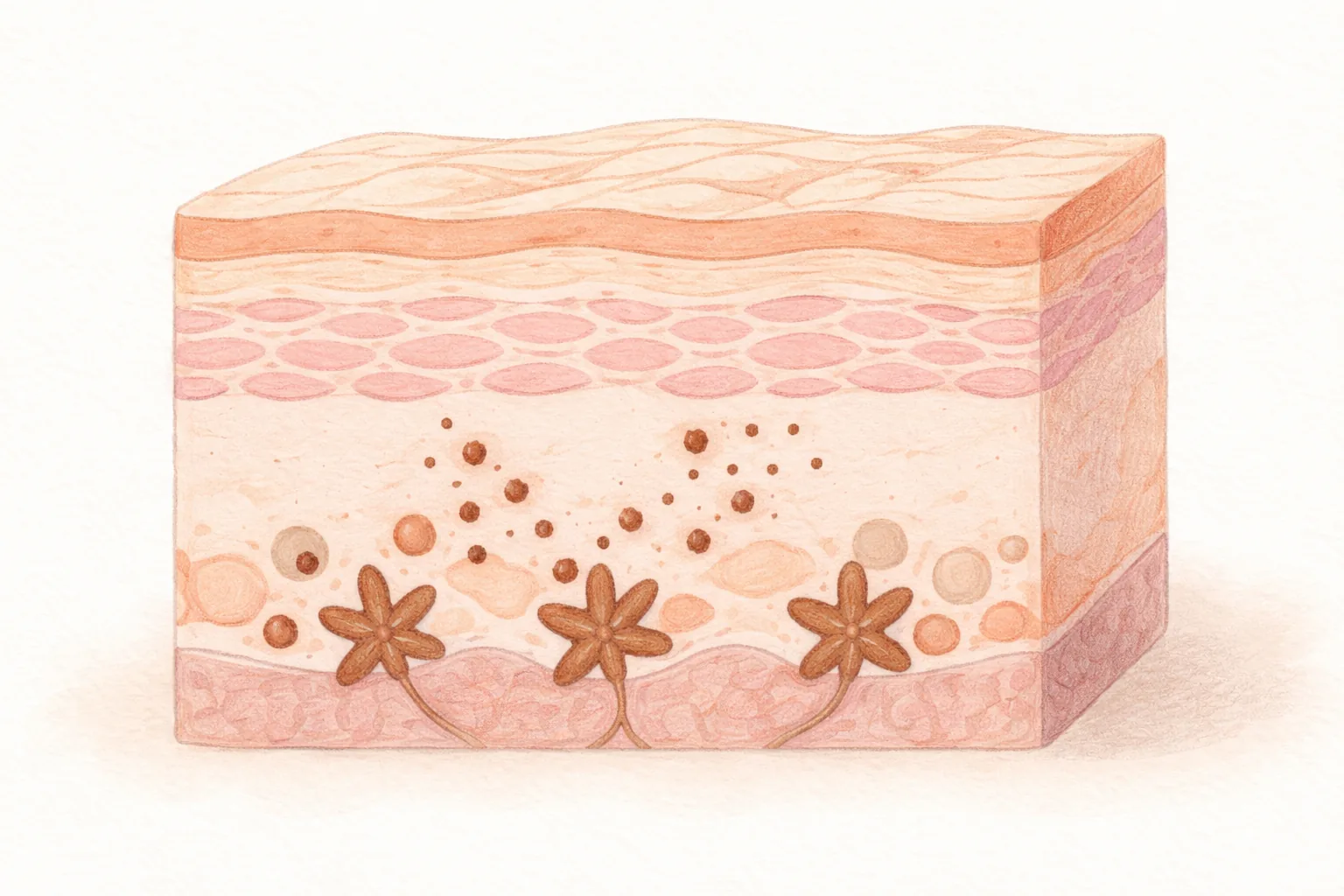
Melanin is the pigment that gives skin, hair and eyes their color, and how much melanin is present determines how pigmented the skin is (MSD Manuals). The cells that make melanin are scattered among other cells in the basal layer, the deepest layer of the epidermis, and the melanin produced there spreads to surrounding cells to create skin color.
The reason the same melanin gets different names is that the type of trigger and where the pigment stays differ. When cumulative UV exposure and aging are the main causes, it appears as relatively well-defined sun spots; when hormones, UV and heat stimulation overlap, it appears as melasma with blurred, widely spread borders.
Freckles have a strong genetic predisposition, appear from childhood, and show a repeating pattern of darkening in summer and fading in winter, which is different in character from sun spots. Seborrheic keratosis becomes more common with age, feels slightly raised to the touch, and can take on a dark brown or black color.
Here is one fork in the road. Brown marks don't only appear because of sun exposure. According to the MSD Manuals, hyperpigmentation can arise through multiple routes, including inflammatory conditions, drugs or substances, healed skin inflammation such as atopic dermatitis or psoriasis, and sun exposure. Even with the same brown color, a different cause means a different approach.
Sharp borders mean sun spots, blurred borders mean melasma — how far does this rule hold?
It's a commonly used rule, but in practice it often breaks down. Melasma usually spreads symmetrically on both cheeks with blurred borders, while sun spots appear scattered on sun-exposed areas and tend to be small. But it's not uncommon for melasma and sun spots to overlap in the same area, or for a form to sit somewhere between freckles and melasma (ATTITUDE Clinic column). That's why it's hard to reach a conclusion at home just by looking at color and location.
Color left after inflammation is another confusing item. According to the American Academy of Dermatology, when a flat colored spot remains where acne has settled, it is more likely post-inflammatory hyperpigmentation than a scar (AAD). A scar leaves a mark even when the color fades, but post-inflammatory hyperpigmentation usually fades over time. If you mistake an acne mark for melasma and target it with a strong pigment laser, it can add irritation.
So what you need before treatment is not naming it but checking how deep the pigment is. Pigment sitting shallow in the epidermis and pigment that has descended into the dermis cannot use the same device with the same settings, and the approach differs. Pigment type is ultimately judged by a doctor through examination.
Why do dark spots and sun spots appear and why do they increase?
Dark spots and sun spots form where cumulative UV exposure meets skin aging. Melanin is produced more to protect the skin when it receives stimuli such as UV, heat, inflammation or hormonal changes, and when this melanin accumulates excessively in a specific area, it settles as a well-defined pigmented spot (ATTITUDE Clinic column).
Even with the same amount of sunlight, the results differ from person to person. Because the amount of melanin produced and the way it spreads differ by person, some people develop melasma, some get more sun spots, and some see no major change. With age, the skin's regenerative capacity declines, making it harder for a spot that has appeared to fade, and without UV protection the number and range gradually increase. The American Academy of Dermatology also notes that how to remove age spots and dark spots is a frequent question for dermatologists, and that effective treatment begins with understanding why the hyperpigmentation occurred (AAD).
One thing must be pointed out. For a mole that is new or growing, or that changes in shape or color, or itches or bleeds, the order is to check for skin cancer through examination before considering cosmetic procedures. Some skin cancers can look like an ordinary brown spot (ATTITUDE Clinic column).
Why do results differ even with the same laser?
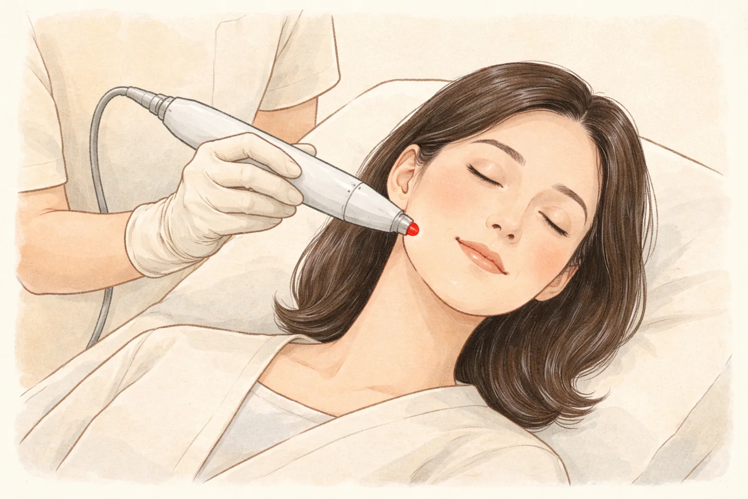
Pigment lasers work by having melanin selectively absorb light of a specific wavelength to break up pigment particles (ATTITUDE Clinic column). Because each wavelength reaches a different depth in the skin, shallow sun spots in the epidermis and melasma that has spread into the dermis are approached differently from the start. This principle of focusing energy on the target while leaving surrounding normal skin less touched is called selective photothermolysis (ATTITUDE Clinic column).
What happens if strong energy is used on melasma? The pigment-producing cells can be stimulated, temporarily appearing lighter before actually darkening — rebound hyperpigmentation. So the basic approach for melasma is to lower the intensity and repeat at intervals, along with UV protection and, when needed, medication (ATTITUDE Clinic column).
How many sessions you need depends on the goal and pigment depth. In the ATTITUDE Clinic column, common examples are given: sun spots get 1–3 sessions of picosecond-type treatment at 4–8 week intervals, while melasma or widespread pigment gets 8–12 or more sessions of low-power toning at least 1 week apart (ATTITUDE Clinic column). These numbers are not a fixed answer but closer to a starting point, and the actual number is adjusted while watching progress.
Making intervals too short is also a problem. Frequent laser toning has been reported in association with mottled hypopigmentation, so toning is usually spaced at least 1 week apart (ATTITUDE Clinic column). Giving the skin time to recover gets closer to results than filling out sessions.
Table 1 Why do results differ even with the same laser?
Pigment type
Layer it sits in
Main approach considered
Chance of returning
Seborrheic keratosis
Epidermis (outer layer thickened)
Removing the thickened area with an erbium:YAG laser, etc.
Low (new ones can appear)
Freckles / sun spots
Epidermal basal layer
Picosecond-type treatment focused on individual spots, with UV protection
Medium
Melasma (epidermal)
Epidermal basal layer
Repeated low-energy pico toning, with medication and sunscreen
High
Melasma (dermal / mixed)
Epidermis to dermis
Long-term low-energy toning and combined approach
High
Swipe sideways to see the whole table.
In summary The real criteria for sorting brown spots are not color but the location and cause of the pigment. Pigment shallow in the epidermis responds relatively quickly, while melasma spread into the dermis is managed with low energy over a long period to reduce irritation. Seborrheic keratosis is not a pigment problem but a matter of treating thickened epidermis, so the approach is separate.
If this applies to you
If you have a fixed date like a wedding or a photo shoot
Recovery time varies greatly depending on the type and intensity of the procedure. Scabs that form after sun spot laser usually fall off in about 7–10 days, so if you have an important event, work backward from the date to set the timing (ATTITUDE Clinic column). If you tell us your schedule at the consultation, we can build a combination that avoids excessive intensity.
If it's a season with a lot of outdoor activity
When UV is strong, the risk of post-treatment hyperpigmentation is higher than in other seasons. On the premise of thorough UV protection, treatment can proceed regardless of season, and if your lifestyle involves a lot of outdoor time, it's better to postpone high-power treatments with long recovery to when exposure decreases. Low-power toning or maintenance treatments have relatively fewer seasonal restrictions.
If redness and sun spots appear together
Even on the same face, sun spots come from pigment, redness from blood vessels, and rough texture from the skin surface (ATTITUDE Clinic column). Because the devices targeting pigment and those targeting blood vessels are different, it's less burdensome to decide the order of what to address first rather than doing it all at once.
If your skin tends to be sensitive
If the energy is too strong for your skin condition or repeated before recovery, redness can linger and the barrier can become sensitive. If cosmetics feel stinging or you've recently had severe redness, that fact is important in setting the parameters.
Tell us before your consultation Your UV exposure over the past few weeks (travel or outdoor activity), medications you're taking and contraceptive or hormonal changes, exfoliants or retinoid products you're using, reactions you've had from previous procedures (bruising, scabs, experience of pigment darkening), and fixed dates like a wedding or a photo shoot. With these five points, we can set the intensity and timing much more specifically.
How ATTITUDE Clinic proceeds
AI skin analysis record — Current skin condition such as pigment, redness, texture and pores is recorded with an analysis tool. This record is a tool for organizing information to help the consultation and is not a medical diagnosis.
Doctor consultation to interpret type and cause — The analysis record is combined with lifestyle information such as UV exposure and hormonal changes and your concerns, and the doctor judges the pigment type directly through examination. You decide together what kind of pigment it is, what is needed and not needed now, and the order.
Doctor-performed treatment — Among the devices held, such as PicoPlus, Vbeam and erbium:YAG laser, the one suited to the pigment type is chosen, and the doctor adjusts intensity and combination. Rather than insisting on one device, the choice matches whether the concern comes from pigment, blood vessels or the skin surface.
Recovery and care guidance — Precautions right after the procedure, UV protection and the timing of the next follow-up are explained. The principle is not to recommend more sessions than necessary, and intensity and intervals are adjusted while watching progress.
Post-treatment care and side effects
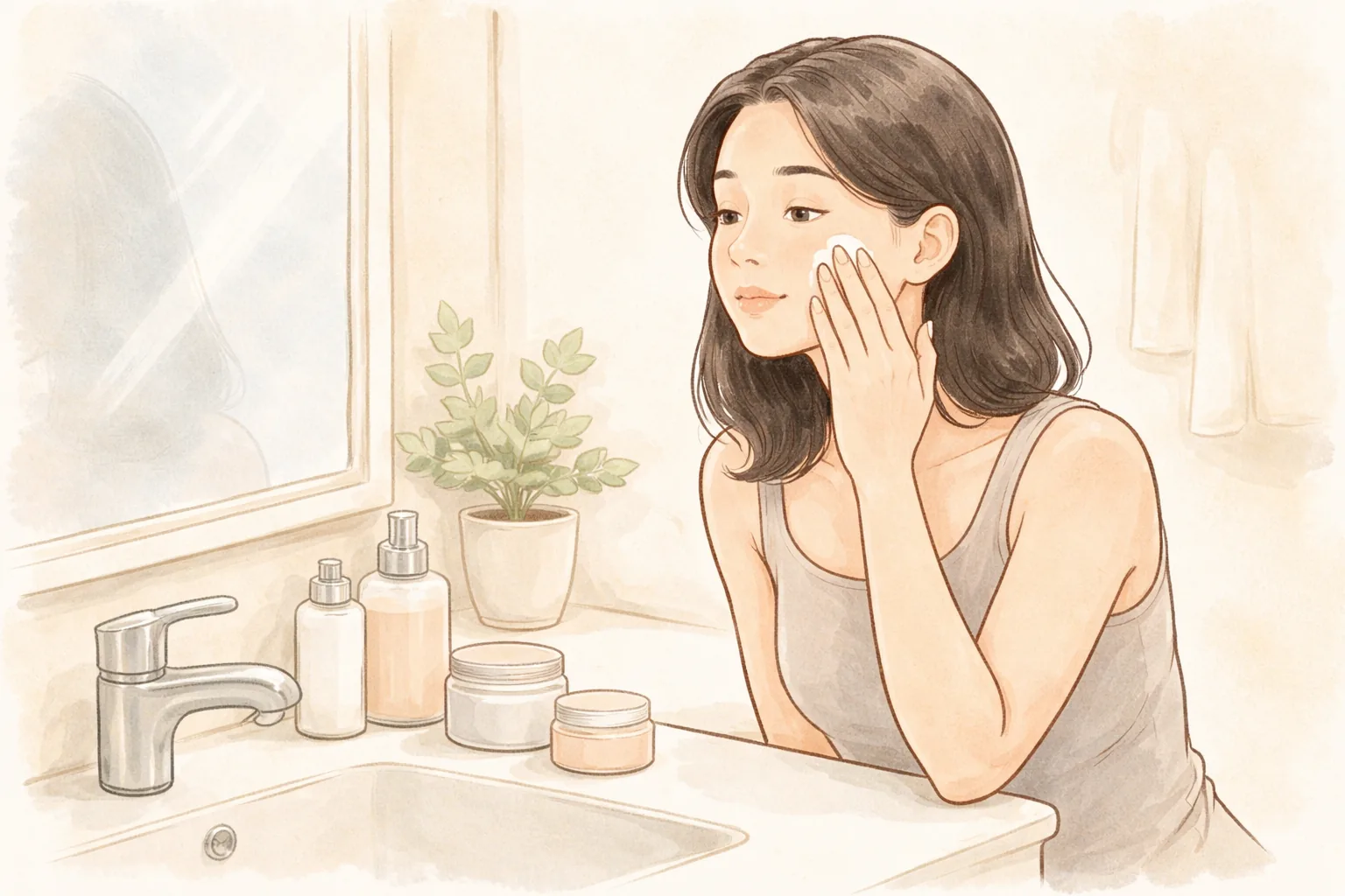
Soothing and moisturizing — Skin temperature is elevated right after the procedure. Wipe lightly with lukewarm rather than hot water without rubbing, and apply a low-irritation moisturizer frequently to help the barrier recover.
UV protection — Apply SPF 50+ sunscreen for at least 2 weeks after the procedure, and reapply about every 2 hours outdoors (ATTITUDE Clinic column). Protection during this period is the main means of reducing pigment recurrence.
Heat stimulation and scab care — Avoid saunas, jjimjilbang and intense exercise for 1–2 weeks. If a scab forms, don't touch it and let it fall off on its own.
Checking progress — If redness or heat lasts longer than expected or pain worsens, don't endure it — come in to have it checked. Even if you feel the pigment is darkening again, don't raise the intensity on your own.
Common reactions are redness, heat, swelling and stinging, and some procedures form scabs. The color of the treated area may temporarily darken, and vascular lasers can cause bruising. Rarely, blisters, hypopigmentation where the skin color becomes lighter, infection, scars, or pigment returning later can occur (ATTITUDE Clinic column). Results and recovery vary by individual, and the doctor explains the side effects of the planned device and settings before the procedure.
Frequently asked questions
How do you tell melasma from sun spots before treatment?
Borders and distribution are the starting point. If it spreads widely and symmetrically with blurred borders, think melasma first; if it's scattered on sun-exposed areas with sharp borders, think sun spots first. However, overlapping cases are not uncommon, so there are limits to judging by eye alone, and pigment type is judged by a doctor through examination (ATTITUDE Clinic column). In consultation, we look at how deep the pigment is, when it appeared, and whether there have been changes such as pregnancy or contraceptive use.
Are seborrheic keratosis and dark spots the same?
They are different. A dark spot is a spot where melanin pigment is locally excessive, while seborrheic keratosis is a benign lesion in which the outer skin cells thicken, with color being a secondary feature (ATTITUDE Clinic column). So seborrheic keratosis is treated by smoothing the raised part rather than by a laser that breaks up pigment. It can feel raised to the touch and take on a dark brown or black color.
How do laser toning and sun spot laser differ?
The goals differ. Sun spot laser focuses on targeting individual spots to break up pigment particles, while toning repeatedly applies low energy to the whole face to slowly calm the overactivity of pigment cells. So for widely spread pigment like melasma, low-power toning, UV protection and medication are the basics (ATTITUDE Clinic column). Even with the same device, the role changes depending on how intensity and treatment range are set.
How many sessions before I see results?
There is a large difference depending on pigment depth and skin condition. Common examples suggest 1–3 sessions at 4–8 week intervals for sun spots, and 8–12 or more sessions at least 1 week apart for melasma or widespread pigment (ATTITUDE Clinic column). Pigment is not completed in 1 session, and the skin needs time to process the broken-down pigment. What matters more than the number of sessions is whether intervals and UV protection are maintained together.
Points to look at when choosing a clinic for pigment treatment
Look at the order, not the device list. First, does the clinic confirm whether your concern is pigment, blood vessels or a skin surface problem, and is that judgment made through examination? Next, does it recommend only the necessary procedures and not fix the number or intensity in advance, and do you hear enough about pain, recovery schedule and post-treatment precautions at the consultation? Finally, is it a place where you can be checked if discomfort arises during recovery?
At a glance
Table 2 At a glance
Item
Key point
Commonality
All look like brown spots, but the causes differ
Biggest difference
The layer the pigment sits in and the cause
Seborrheic keratosis
Not pigment but a benign lesion of thickened epidermis
Difficult pigment
Melasma: strong energy backfires, low-energy repetition is the basics
Treatment direction
Design wavelength and intensity to match pigment type and depth
This article offers general health information. It is not a substitute for diagnosis or treatment based on your own condition, so please talk through your situation with a doctor.