Pico Laser for Melasma: Why Boundaries and Depth Determine Your Treatment Plan
Why melasma, sun spots, and seborrheic keratosis look the same brown—and how boundaries and depth determine the treatment order when they appear together.
Written by ATTITUDE Clinic Editorial Team
When you look in the mirror, it's common to wonder whether the brown patches on your cheeks are melasma, the dot-like solar lentigines, or raised seborrheic keratosis. All three appear brown, but they arise from different mechanisms and sit at different depths, so the treatment approach differs. The order in which you read the boundaries and depth—and what to address first when they coexist—determines the actual treatment.
The Bottom Line First
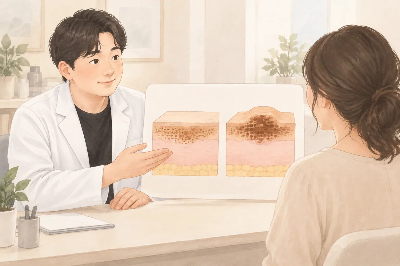
Melasma has blurry borders and spreads widely, with pigment possibly spanning from the epidermis to the shallow dermis. Solar lentigines and seborrheic keratosis usually have distinct borders and sit in the epidermis, but seborrheic keratosis is a benign lesion with thickened cells of the outer skin layer rather than simply accumulated pigment. Therefore, for solar lentigines and seborrheic keratosis, the basic approach is to target and remove the lesion directly, while for melasma, the basic approach is to use low energy repeatedly to stabilize pigment cell activity. Since they can coexist on one face, it is safer to first check boundaries and depth, and then divide the intensity of stimulation and recovery intervals for each lesion.
Why do melasma, seborrheic keratosis, and solar lentigines all look the same brown?
The cells that produce skin color are called melanocytes (cells that make skin pigment). Melasma, freckles, and spots all start from these cells, but seborrheic keratosis is a benign lesion where epidermal cells thicken, so the principle is different. ATTITUDE Clinic's column also highlights this difference first.
Melanin is produced more to protect the skin when stimulated by UV rays, heat, inflammation, or hormonal changes. According to the American Academy of Dermatology, UV rays can make the skin produce more pigment, darkening existing melasma and creating new spots (AAD). Melasma is a condition where pigment cells are overactivated by a combination of hormonal, UV, and heat triggers, with unclear borders that tend to spread widely. Solar lentigines are relatively distinct brown spots caused by cumulative UV exposure and aging, and seborrheic keratosis may feel slightly raised to the touch.
What matters more than the shared brown color is which layer the pigment sits in and by what mechanism it developed. So naming it based on color alone can lead to inconsistent responses even with the same laser. Any new or growing mole, or one that changes in shape or color or bleeds, should be checked by a doctor before cosmetic treatment.
Can you just say distinct borders mean solar lentigines and blurry borders mean melasma?
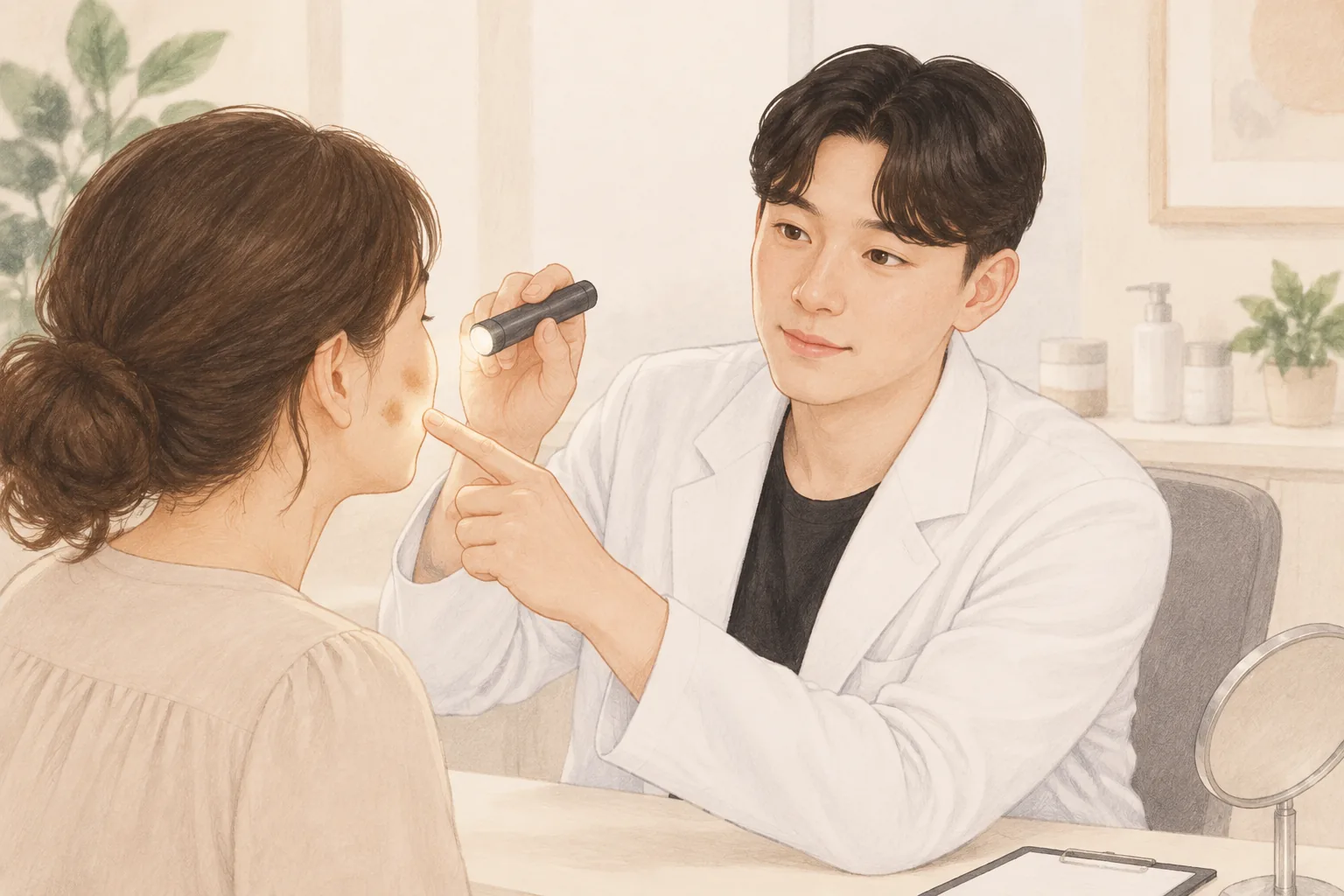
Borders are a good clue, but they aren't the whole story. This is why we use a Wood's lamp (a diagnostic tool that illuminates pigment depth) or a dermoscope (a device that magnifies the skin) to see how deep the pigment reaches. The AAD notes that melasma can look like other skin conditions, and if needed, a skin biopsy can help differentiate (AAD).
Melasma is also divided into epidermal, dermal, and mixed types, and even within the same melasma, the depth of pigment involvement varies. Spots grouped as solar lentigines or blemishes often include lesions like solar lentigines that sit relatively shallow in the epidermis, but when solar lentigines overlap on a melasma background, it's hard to distinguish them by borders alone. This is why it's difficult to accurately differentiate at home based only on color and location.
More than whether the borders are distinct, the depth the pigment reaches determines the next treatment. Even a small spot, if it has changed recently, requires examination before pigment treatment.
When melasma and solar lentigines coexist, how is the treatment order decided?
Melasma, solar lentigines, and freckles can all appear on the same face. It's not uncommon to see distinct-bordered spots overlapping on a melasma background, or forms somewhere between freckles and melasma. Therefore, rather than concluding with one name, we check the boundaries and depth of each lesion.
Seborrheic keratosis is a benign lesion with thickened epidermal cells, so removal of the raised portion with an erbium YAG laser or similar is common. For melasma, the basic approach is repeated management with low-energy toning to avoid stimulating pigment cell activity. When both are present, we don't treat all lesions the same way on the same day; we divide the order considering the stimulation and recovery interval for each lesion.
Rather than trying to erase everything at once because they coexist, we divide the intensity of stimulation and recovery interval for each lesion. If the skin barrier is weakened, strong stimulation may be postponed.
Table 1 When melasma and solar lentigines coexist, how is the treatment order decided?
Pigment Type
Layer
Laser Approach
Recurrence Potential
Seborrheic keratosis
Epidermis, thickened outer layer
Removal with erbium YAG laser, etc.
Rare, may develop new ones
Solar lentigines/blemishes
Mainly basal epidermis
Individual spot approach with pico spot, etc.
Moderate
Epidermal melasma
Basal epidermis
Repeated low-energy pico toning, concurrent sunscreen and medication
High
Dermal/mixed melasma
Epidermis to dermis
Long-term low-energy management and combined approach
High
Swipe sideways to see the whole table.
In summary Seborrheic keratosis is a lesion with thickened cells, solar lentigines are distinct-bordered epidermal pigment, and melasma is a widely spreading problem of overactive pigment. When they coexist, dividing the order after checking each lesion's depth and stimulation reduces the recovery burden.
Why shouldn't lasers be used at the same intensity?
Laser treatment uses light of a single wavelength absorbed by a target, converting it into heat or shock. This principle is called selective photothermolysis (targeting energy to the target while minimizing impact on surrounding areas). Each wavelength reaches a different depth in the skin, and the intensity of energy affects the stimulation to the skin.
Picosecond pulses are as short as one trillionth of a second, breaking pigment particles into fine fragments. On the other hand, using strong energy on melasma can cause temporary lightening followed by rebound hyperpigmentation as pigment cells are stimulated and darken. The blemish, redness, and scar laser guide also cites this as a reason to distinguish melasma from blemishes.
For melasma treatment, laser toning with low output delivered over multiple sessions is widely used. A PMC review explains that laser toning is used as a safe and effective method for melasma management, and satisfactory responses are often seen when combined with topical agents. A network meta-analysis also showed that Q-switched Nd:YAG laser combined with topical agents ranked highly, but notes that the actual choice should consider side effects, skin type, and lesion duration.
For melasma, repeated low-energy sessions reduce stimulation better than one strong session. Using strong energy on melasma may cause temporary lightening but can lead to darkening.
If this applies to you
When a dot-like brown spot suddenly appears
A small, distinct-bordered spot may be a solar lentigo, but if it appeared recently or is changing in size or color, an examination is needed first. Before cosmetic treatment, ruling out skin cancer is the first step. Even if it seems unchanged, if it itches or bleeds, the same order applies.
When seborrheic keratosis appears to overlap on a melasma background
When raised seborrheic keratosis overlaps on melasma, the urge to clear everything at once is strong. However, each lesion sits at a different layer and responds differently to stimulation, so which lesion to address first is decided during consultation. We avoid concentrating strong energy on melasma areas.
When a wedding or photo shoot is coming up
After pigment treatment, redness or crusting may occur, so the interval before your event matters. Recovery time varies by lesion type and treatment intensity, so letting us know your dates in advance helps adjust intensity and timing. For first-time treatments, start with more buffer than right before an event.
When your skin is sensitive or prone to redness
If the skin barrier is weakened, we first assess inflammation, redness, and dryness before pigment treatment. Even for the same pigment, we can start with a less irritating approach, and if recovery is slow, we lengthen the interval. If your face easily flushes from saunas or exercise, mention this during consultation.
Please tell us before consultation Recent UV exposure, medications taken or applied, whitening or exfoliating products, previous experiences of sensitivity after laser or peeling, and dates for weddings, photo shoots, or travel. If you have any new or changed moles, please mention them as well.
How ATTITUDE Clinic Proceeds
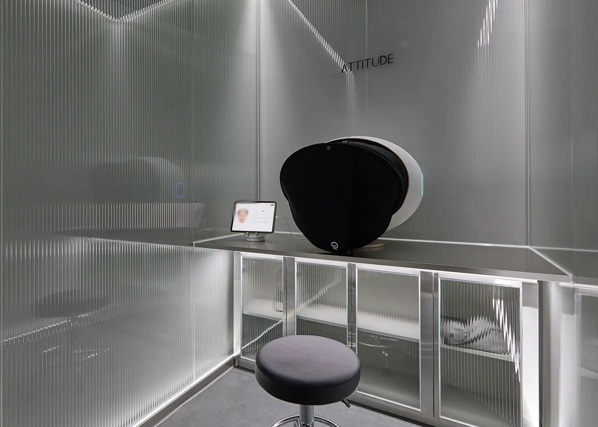
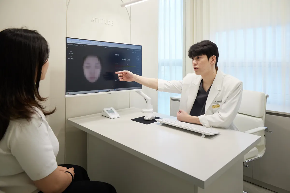
Precision diagnosis and skin analysis We record your skin condition with devices and organize pigment distribution, redness, and texture together. The analysis tools are used to record and organize information and do not make medical diagnoses.
Doctor consultation and type assessment The doctor adds UV exposure, hormonal changes, and previous treatment experience to the analysis records and determines the pigment type through examination. We decide together which lesion to address first and what treatment is needed now versus not needed.
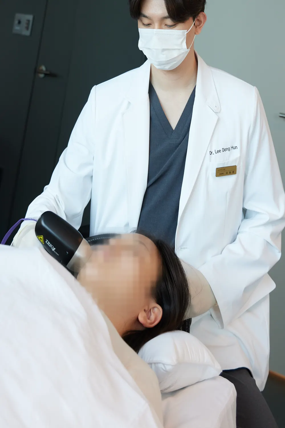
Doctor-performed treatment Based on the pigment type, the doctor selects the necessary device from our available equipment such as PicoPlus, Vbeam, and erbium YAG laser, and adjusts intensity and combination directly. We proceed only as much as needed to avoid excessive stimulation.
Four steps your doctor sees to in person
From the diagnostic device to the treatment bed, the doctor you first consult takes care of everything.
01
Precision diagnosis
In a private diagnosis room the whole face is photographed, recording even skin conditions that are hard to see with the eye.
02
Skin analysis
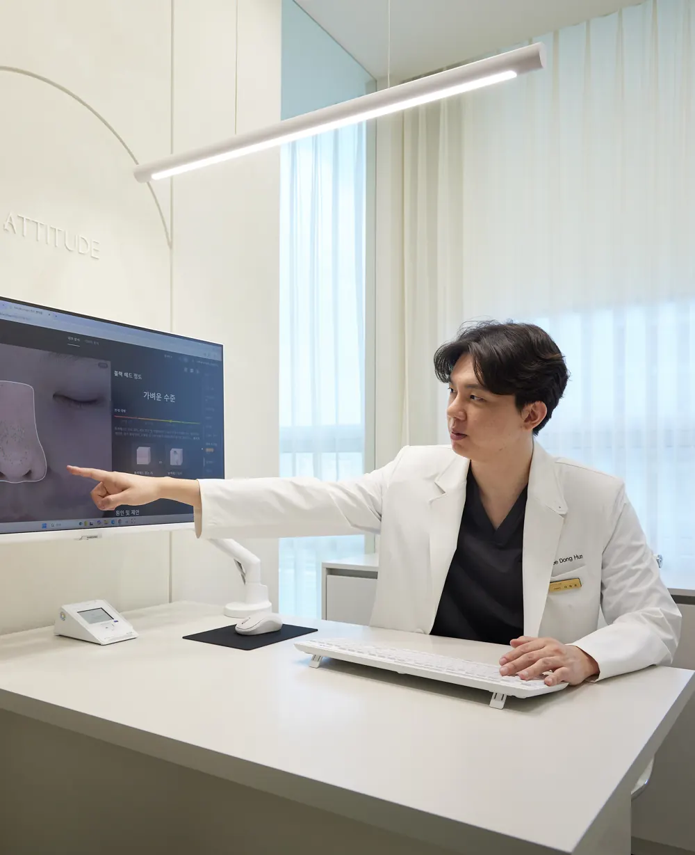
The doctor goes through your pore, pigmentation, wrinkle and firmness results with you on screen and explains what comes first.
03
One-to-one design
With your skin, the balance of your face, the change you want and your schedule in view, you decide the treatment and its extent together.
04
Treatment by your doctor
The doctor who consulted you performs the treatment from start to finish in a private treatment room. No one takes over halfway.
“I take injection pain seriously.”
Co-Director Dr. Lee Dong Hun
Many people hesitate over injection treatments because they hurt. So we went back over everything, from the anesthetic to the products used.
Careful numbing, unhurried timingWe allow plenty of time for a special numbing cream to take full effect, and add anesthetic injections for the treatments and areas that need them.
Fine needles, finished quicklyInjection treatments use fine needles, and the doctor finishes them personally in a short time, adjusting along the way by watching how you are doing.
Even the product formulation countsThe same treatment can feel more or less demanding depending on the product, so we take that into account too.
The anesthetic method and whether it is used are decided after an examination, according to the treatment, the area and your condition. How much it hurts varies from person to person.
Post-Treatment Care and Side Effects
Soothing Redness and heat may occur right after treatment, so gently wipe with lukewarm water without rubbing.
Moisturizing While the skin barrier is weakened, apply a mild moisturizer thinly multiple times.
UV protection The AAD recommends using broad-spectrum sunscreen along with hats and shade (AAD). After pigment treatment, follow clinic instructions to reapply SPF 50+ every 2 hours for 2 weeks.
Follow-up Crusts often fall off on their own within 7–10 days, so do not pick them. If redness or stinging lasts more than 3 days or suddenly worsens, contact the clinic without waiting.
Commonly, the treated area may become red or swollen, crust may form, or color may temporarily darken. Rarely, hypopigmentation (lightened skin), blisters, or scars may occur, and pigment may return over time. Recovery speed and final color depend on pigment depth, skin thickness, and UV exposure.
Frequently Asked Questions
Can melasma and solar lentigines coexist?
They can coexist on one face. It's not uncommon for distinct-bordered solar lentigines or freckles to overlap on a melasma background. Therefore, we don't conclude with one pigment type but check the boundaries and depth of each lesion. Treatment doesn't treat all lesions the same way on the same day; we divide the order considering stimulation and recovery intervals. We don't perform strong treatments first just because they coexist.
A dot-like brown spot—is it a solar lentigo or melasma?
If it has distinct borders and looks like a small dot, it may be a solar lentigo; if it's blurry and spreads widely, it's more likely melasma. However, there are many intermediate forms, making it hard to distinguish by eye alone. Melasma also has epidermal, dermal, and mixed types that can look similar. This is why we examine the depth of pigment during examination. During consultation, we use a Wood's lamp or dermoscope to check how deep the pigment reaches, and if needed, a biopsy to differentiate. If a spot is newly appeared or changing in size and color, examination is recommended before pigment treatment.
When seborrheic keratosis and melasma coexist, what is treated first?
Seborrheic keratosis is a lesion with thickened epidermal cells, so removal with an erbium YAG laser is common, while melasma is managed with repeated low-energy toning. When they coexist, the order is decided considering each lesion's stimulation and recovery interval. If the skin barrier and inflammation are not good, we address that first. What to do first is decided after examination.
Should melasma not be treated with lasers?
It's not a contraindication but a matter of intensity and interval. Using strong energy on melasma can stimulate pigment cells, causing temporary lightening followed by darkening. Therefore, the basics are repeated low-output laser toning, UV protection, and concurrent topical agents. Results can be expected when combined with cause management rather than laser alone.
What should I look for when choosing a clinic for pigment treatment?
Look for whether they first check the boundaries and depth of your pigment rather than equipment names. Also check whether they explain the treatment order for each lesion, discuss recovery time and side effects during consultation, and whether you can follow up if you have discomfort after treatment. AI skin analysis is a tool to help record and organize, and pigment type assessment is made by the doctor's examination. Recovery speed and recurrence vary with skin condition and lifestyle.
At a Glance
Table 2 At a Glance
Item
Key Point
Commonality
All look like brown spots
Biggest difference
The layer where pigment sits and the mechanism
Seborrheic keratosis
Benign lesion with thickened epidermal cells, removal approach
Solar lentigines/blemishes
Distinct borders, mainly epidermis, target the lesion
This article offers general health information. It is not a substitute for diagnosis or treatment based on your own condition, so please talk through your situation with a doctor.